


Cell-level maps released
 Print
Print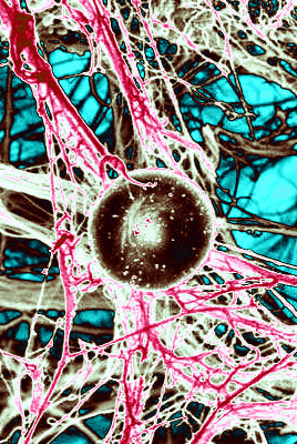 Researchers have produced detailed maps of all the cells in human organs.
By studying how trillions of cells interact, connect, and form tissues, the maps promise to revolutionise understanding of health and disease.
Published as part of the Human BioMolecular Atlas Program (HuBMAP), three new papers present reference cell maps for the human intestine, kidney, and maternal-foetal interface.
These maps provide invaluable insights into how cell types are arranged and interact within different human tissues and organs.
The Human BioMolecular Atlas Program (HuBMAP) is an ambitious initiative with the aim of comprehensively mapping cell arrangements throughout the human body.
By doing so, scientists hope to gain new knowledge about cellular functions and their impact on an individual's health.
The HuBMAP consortium has been developing advanced tools capable of assembling spatial maps of cell molecular components such as RNAs, proteins, and metabolites at a single-cell level.
In a study of the human intestine, researchers Michael Snyder, Garry Nolan, William Greenleaf, and their team analysed eight sections of the intestine from nine individuals.
The findings revealed striking variations in cell composition across different regions. Notably, they identified previously unknown subtypes of epithelial cells that play crucial roles in immune responses, shedding light on the intricate mechanisms governing this vital organ's functioning.
Researchers Sanjay Jain, Matthias Kretzler, Kun Zhang, Tarek M El-Achkar, Pierre C Dagher, and Michal T Eadon focused on the human kidneys, analysing cells from 45 healthy and 48 diseased kidneys.
By producing a single-cell and spatial atlas of 51 main cell types across different regions of the kidney, they uncovered cellular states and neighbourhoods affected by acute or chronic injury.
This breakthrough has significant implications for understanding kidney function and developing targeted treatments.
Another team led by Michael Angelo and Shirley Greenbaum constructed a comprehensive map of the human placenta during the first half of pregnancy.
By analysing around 500,000 cells and 588 arteries from 66 samples of the maternal-foetal interface, they examined the dynamic interactions between placental and immune cells.
This discovery has profound implications for understanding how maternal immune cells support the coexistence of maternal and foetal cells, ultimately ensuring a healthy pregnancy.
The full collection will be available upon publication here.