


Immune cell 'gulps' spotted
 Print
Print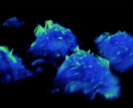 Scientists have, for the first time, watched in real-time as immune cells survey their surroundings to detect threats.
Macrophages are a type of white blood cell whose job it is to detect and engulf any material that may be a potential danger to our health – such as debris of old cells, microbes, cancer cells and foreign substances.
They survey the body by taking in large gulps of surrounding fluid to identify anything they do not recognise as healthy, triggering immune responses when necessary to ward off infection.
Scientists at the University of Queensland have been able to record the gulping mechanism, known as macropinocytosis, in real time on live cells using a cutting-edge laser microscope called a lattice light sheet, which captures huge 3D images with great precision in just seconds.
The team discovered new structures they termed ‘tent-pole ruffles’ on the surface of immune cells, which help the cells gulp the surrounding fluid for sampling.
The new technique redefines scientists' understanding of the macropinocytosis process, and immune function and more broadly, Professor Jenny Stow says.
“This imaging will give us phenomenal power to reveal how cell behaviour is affected in disease, to test the effects of drugs on cells, and to give us insights that will be important for devising new treatments,” she said.
In cancer, for instance, the process of macropinocytosis is needed by aggressive cancer cells to gulp in the nutrients they need to sustain growth.
Cancer cells also form ‘tent-pole ruffles’ and the scientists are now pursuing molecules they have identified on the tent-pole ruffles as targets for blocking cancer cell survival.